Table of Contents
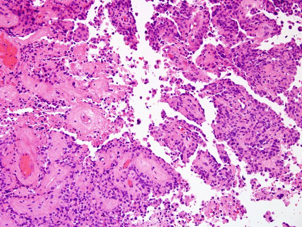
Washington University Experience | NEOPLASMS (GLIAL) | Astroblastoma | 5B2 Astroblastoma, anaplastic (Case 5) H&E 18.jpg
5B2-7 Sections of the solid portion of the resected tumor show sheets of tumor cells separated by numerous thick walled small to moderately sized vessels, which focally imparts it a papillary to pseudo-papillary architecture. Geographic tumor necrosis without pseudopalisading is multifocal. Tumor nuclei are closely juxtaposed to the vessel walls with a pseudo-rosetted appearance. The cytoplasmic background is however not very fibrillary and instead tumor cells have prominent cell borders and epithelioid morphology. Additional cytologic features include abundant glassy eosinophilic cytoplasm with eccentric nuclei, multinucleation and moderate to focally severe nuclear pleomorphism. There are up to 12 mitotic figures in 10 high-power fields and atypical forms are present. Tumor is seen infiltrating into the overlying dura.

