Table of Contents
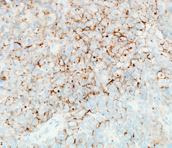
Washington University Experience | NEOPLASMS (GLIAL) | Astroblastoma | 5E2 Astroblastoma, anaplastic (Case 5) D2-40 1.jpg
D2-40 immuno-labeling shows strong membranous positivity in a subset of cells as well as fewer cells with perinuclear dot-like staining. ---- Other (not shown): Neoplastic cells are negative for CD34 and reticulin content is mostly low in the tumor. Both CD34 and reticulin however highlight its highly vascular nature. INI1 shows retained nuclear expression. There is no immunoreactivity for mutant IDH1 (R132H). Ki-67 shows high proliferation rate (up to ~23.8%). BRAF (V600E) immunostain interpretation is equivocal due to its high background. Trichrome demonstrates hyalinized vessels including smaller sized vessels within the tumor. ---- Comment: The collective findings are those of a high-grade glioma, and our main differential considerations included anaplastic ependymoma and anaplastic (malignant) astroblastoma, the latter which we favor because of its overall very epithelioid appearance and lack of fibrillary quality in most parts. Even though both the tumors can demonstrate focal epithelial membrane antigen expression, GFAP immunostain in this particular case lacks the fibrillary background and thin perpendicularly oriented perivascular processes that are more characteristic of anaplastic ependymoma.

