Table of Contents
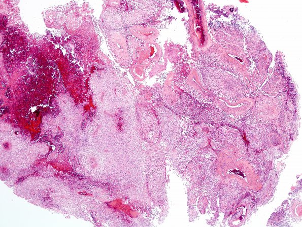
Washington University Experience | NEOPLASMS (GLIAL) | Astroblastoma | 7A1 Astroblastoma, anaplastic (Case 7) H&E 3.jpg
Case 7 History ---- The patient is a 58-year old woman with a right parietal mass. ---- 7A1-7 Sections reveal a moderately cellular glial neoplasm with mild to moderate nuclear pleomorphism. The majority of tumor cells have oval nuclei with occasional prominent nucleoli. The tumor cells range from gemistocyte-like to epithelioid and much of the tumor has a solid growth pattern. At the edges though, infiltration of adjacent brain parenchyma is evident. In some areas, there are very prominent perivascular pseudorosettes with broad processes or columnar epithelioid cells arranged around central blood vessels. Other areas have markedly hyalinized blood vessels. The mitotic index is focally elevated. Endothelial hyperplasia and focal necrosis are both present.

