Table of Contents
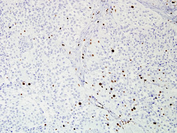
Washington University Experience | NEOPLASMS (GLIAL) | Astroblastoma | 7C Astroblastoma, anaplastic (Case 7) Ki67 2.jpg
An MIB-1 (Ki-67) stain reveals a low proliferative labeling index. ---- Other immunohistochemistry (not shown) GFAP was strong with widespread immunoreactivity. Only rare p53 positive cells are seen. A stain for CD99 highlights a subset of the tumor cells with a membrane staining pattern. ---- The morphologic and immunohistochemical features are consistent with the diagnosis of anaplastic astroblastoma. FISH) was utilized with probes against 1p, 1q, 19p and 19q. These studies showed evidence of polysomes (gains) of both chromosomes 1 and 19. No deletions were identified.

