Table of Contents
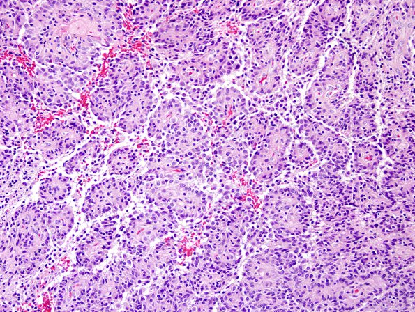
Washington University Experience | NEOPLASMS (GLIAL) | Astroblastoma | 8A1 Astroblastoma, anaplastic vs GBM (Case 8) H&E 4.jpg
Case 8 History ---- The patient is a 44 year old woman who presented with new-onset seizures characterized by the onset of a "vinyl"-like odor in conjunction with lapses of memory and sensation of deja vu. Her symptoms continued to occur two to three times per week associated with headaches, nausea and vomiting. She attributed her symptoms to a recent concussion; a CT scan performed at that time revealed no evidence of mass. However, the patient sought further medical attention when she sustained a neck injury in a motor vehicle accident shortly thereafter. At that time an MRI revealed a contrast-enhancing, lobulated lesion of the superior right medial temporal lobe without evidence of vasogenic edema with mild mass effect and invagination into the basal ganglia. The patient self-referred to a different institution, where repeat imaging performed again showed a well-defined, intensely enhancing, 3 x 2.5 x 2 cm tumor in the right temporal lobe stem without significant surrounding edema or mass effect. The patient underwent subtotal resection of the mass at that OSH. The patient then presented to BJH for further treatment. ---- 8A1-6 H&E stained sections showed multiple fragments of a tumor with predominantly a perivascular pseudo-rosetted architecture; the tumor appears to be deceptively well-circumscribed on low-power in some fragments but has evidence of an infiltrative growth pattern on closer examination; an impression which is also supported by the presence of numerous entrapped axons on a neurofilament immunostain. The cells themselves are rather 'epithelioid' in appearance and are characterized by moderate to abundant pale eosinophilic cytoplasm with stouter cytoplasmic processes extending to the vessels; the nuclei are round-to-ovoid and display mild to moderate pleomorphism with occasional nucleoli. Mitoses are readily identified (up to ~11 per 10 HPF). Minute foci of necrosis and focal hint of incipient microvascular proliferation are additional notable findings.

