Table of Contents
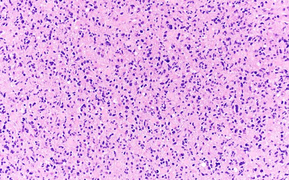
Washington University Experience | NEOPLASMS (GLIAL) | Astrocytoma, IDH-mutant, WHO Grade 4 | 10A1 IDH mut Astro Gr4 (Case 10) H&E 2
Case 10 History ---- The patient was a 41 year old man with a prior diagnosis of oligoastrocytoma, grade II and III on re-resection, MGMT methylated. The most recent MRI showed a large enhancing mass in the same right frontoparietal region at the prior biopsy/resection site. The tumor now involves the corpus callosum. The slides from 2016 resection material were provided for our review. ---- 10A1-3 H&E shows brain parenchyma infiltrated at variable density by a diffuse glioma with regionally heterogeneous cytological features. In some areas, most of the neoplastic cells have oligodendroglial morphology and scattered mini-gemistocytes. In other areas, there are scattered moderately atypical oval nuclei, dense or relatively lucent coarsely speckled chromatin. Mitotic figures are frequent, ranging up to at least 11/10HPF. Some small, thin-walled vascular clusters as well as incipient endothelial proliferation are noted.

