Table of Contents
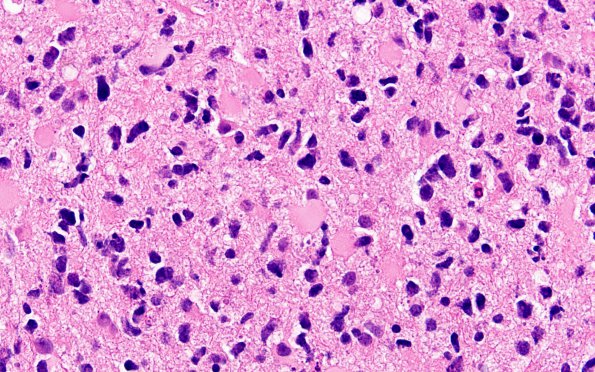
Washington University Experience | NEOPLASMS (GLIAL) | Astrocytoma, IDH-mutant, WHO Grade 4 | 10A2 IDH mut Astro Gr4 (Case 10) H&E 60X 2
This higher magnification image shows brain parenchyma infiltrated at variable density by a diffuse glioma with regionally heterogeneous cytological features. In some areas, most of the neoplastic cells have oligodendroglial morphology and scattered mini-gemistocytes and larger gemistocytes. Mitotic figures are frequent, ranging up to at least 11/10HPF. Some small, thin-walled vascular clusters as well as incipient endothelial proliferation are noted.

