Table of Contents
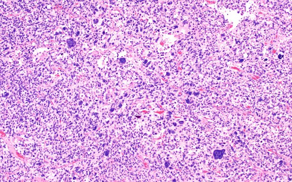
Washington University Experience | NEOPLASMS (GLIAL) | Astrocytoma, IDH-mutant, WHO Grade 4 | 12A1 IDH mut Astro Gr4 (Case 12) H&E 10X
Case12 History ---- The patient is a 35-year-old female who developed headaches, confusion and memory loss. Brain imaging showed a large right frontotemporal mass with edema and subfalcine and uncal herniation. She underwent biopsy and two resections in 04/2015 at an OSH, followed by radiation and Temodar chemotherapy. ---- 12A1-3 H&E stained sections of a histologically heterogeneous high grade diffuse glioma. Many areas are densely populated by tumor cells with small, atypical hyperchromatic nuclei and minimal cytoplasm, accompanied by rare or occasional larger, markedly atypical forms which are numerous in some foci. The smaller tumor cells show more primitive features, with greater hyperchromasia, chromatin smudging, and minimal nuclear molding, somewhat reminiscent of a primitive neuronal component. Other areas are rich in gemistocytic cells, including mucinous microcystic change. Microvascular proliferation is widespread and robust, focally forming garlands and glomeruloid arrangements. Intravascular thrombosis and regions of infarct-like necrosis are also observed.

