Table of Contents
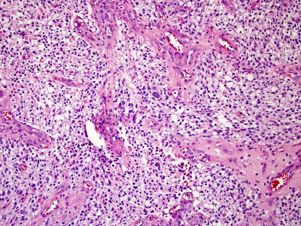
Washington University Experience | NEOPLASMS (GLIAL) | Astrocytoma, IDH-mutant, WHO Grade 4 | 8A1 IDHmut Astro Gr4 (Case 8) H&E 1.jpg
Case 8 History ---- The patient was a 78 year old man with a recurrent meningioma who developed new onset seizures. MRI from 11/2015 showed a second cystic 5.2 cm left frontal lobe lesion lateral to the meningioma that exerted significant mass effect on the left lateral ventricle and intra-operative evidence of invasion into the superior sagittal sinus. Operative procedure: excision. ---- 8A1-3 Sections of the resected left brain tumor show a malignant primary glial neoplasm with several appearances. The majority of the tumor is made up of relatively small, mildly pleomorphic cells diffusely infiltrating the parenchyma. A second population shows greater pleomorphism, cytoplasmic clearing and a more nested appearance due to prolific microvascular hyperplasia and multiple endothelial mitoses. There are large areas of geographic necrosis associated with vascular hyalinization.

