Table of Contents
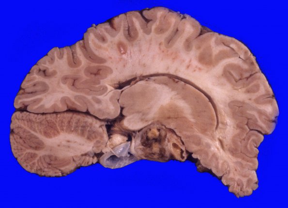
Washington University Experience | NEOPLASMS (GLIAL) | Astrocytoma, pilocytic - Gross Pathology | 2A1 Astrocytoma, pilocytic (Case 2) A1
2A1-4 At autopsy the cerebral hemispheres were symmetrical with gyri and sulci of normal size, shape and distribution. There was no cingulate gyrus, uncal, or cerebellar tonsillar herniation. A 3.3 x 2.4 x 1.8 cm fleshy mass with multiple cysts was present over the hypothalamus which had a central cavity with a yellow rim and was filled with gelatinous grey fluid. It extended into the adjacent thalamus and midbrain and internal capsule. The neoplasm infiltrated into the adjacent cortex and is also noted extending along the Virchow-Robin spaces in the hypothalamus and internal capsule. The pituitary was also invaded by neoplasm.

