Table of Contents
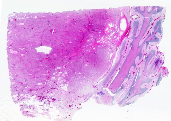
Washington University Experience | NEOPLASMS (GLIAL) | Astrocytoma, pilocytic - Gross Pathology | 6b1 (Case 6) H&E scan WM
Sections of the biopsy and resection specimen show a pilocytic astrocytoma. The tumor is composed predominantly of bland, piloid astrocytes with microcyst formation. Numerous Rosenthal fibers and eosinophilic granular bodies are present. The tumor forms a well-demarcated nodule, abutting the non-neoplastic cerebellum ---- 6B1,2 The microscopic appearance of the specimen shown in 6A illustrating the sharp border (H&E)

