Table of Contents
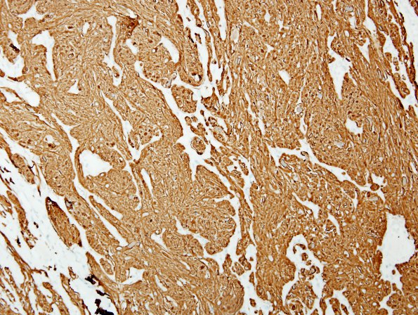
Washington University Experience | NEOPLASMS (GLIAL) | Astrocytoma, pilocytic - Gross Pathology | 7E Astrocytoma, pilocytic, optic nerve (Case 7) GFAP 2.jpg Image id: 25093
GFAP is prominently positive, both in the central portion of optic nerve and in the surrounding tumor. (GFAP IHC) ---- Other findings in this case include: Neurofilament immunostain highlights the nerve axons in the central portion of the optic nerve, but is essentially negative in the surrounding tumor. Epithelial membrane antigen highlights the arachnoid portion of the optic nerve sheath but is negative in the tumor. Ki-67 confirms a low proliferative index in the tumor, highlighting 2-5% of the cells in the various regions of the tumor. P53 highlights only rare positive cells, and accordingly, does not delineate the central portion of optic nerve from the tumor around it. A trichrome stain nicely highlights the Rosenthal fibers (maroon-colored). The retina exhibits ganglion cell loss, correlating with the optic nerve atrophy. Several melanocytic nevi are seen in the iris, involving a variable degree of iris thickness, with some being superficial, while at least one appears to involve the full thickness of the iris stroma, consistent with the clinical history of Lisch nodules.

