Table of Contents
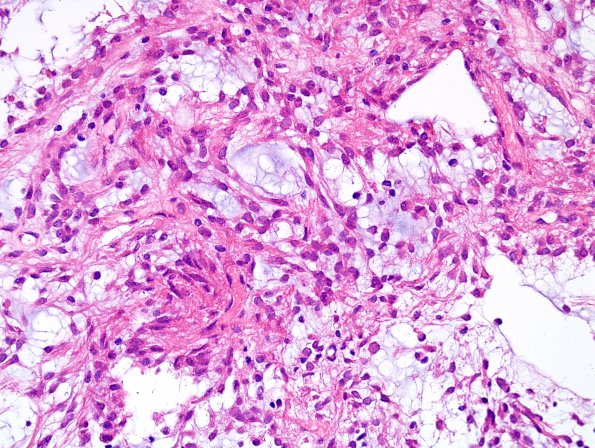
Washington University Experience | NEOPLASMS (GLIAL) | Astrocytoma, pilomyxoid | Filename: 1A2 Astrocytoma, pilomyxoid (Case 1) H&E 10 a (9).jpg
Sections reveal a moderately cellular relatively solid appearing neoplasm with prominent extracellular mucin formation. The pattern was dominated by perivascular pseudorosette-like structures. Endothelial prominence is seen in many of the blood vessels, although well-formed microvascular proliferation is not found. No definite Rosenthal fibers or eosinophilic granular bodies are seen and there is no tumor necrosis. There is mild nuclear pleomorphism, with the majority of tumor cells containing small oval to elongate nuclei with thin eosinophilic cytoplasmic processes. There are prominent small piloid elements in microcystic regions and clumps of astrocytic elements, some with gemistocytic cytoplasm or occasional cells with minimal cytoplasm, suggesting a somewhat more primitive element focally. (H&E)

