Table of Contents
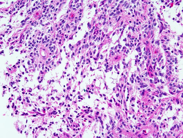
Washington University Experience | NEOPLASMS (GLIAL) | Astrocytoma, pilomyxoid | 3B1 Astrocytoma, pilomyxoid (Case 3) 3.jpg
3B1-4 Sections reveal a moderately cellular, relatively solid-appearing glial neoplasm with a mucin-rich background. There is mild nuclear pleomorphism, with the majority of tumor nuclei being oval with bland chromatin. Most tumor cells have thin hair-like or piloid cytoplasmic processes, some of which are arranged around blood vessels to form perivascular pseudorosettes. No definite Rosenthal fibers or eosinophilic granular bodies are seen on routine sections. There are scattered mitotic figures, but no evidence of endothelial hyperplasia or necrosis.

