Table of Contents
Washington University Experience | NEOPLASMS (GLIAL) | Astrocytoma, pilomyxoid | 4B4 Astrocytoma, pilomyxoid (Case 4) H&E 11.jpg
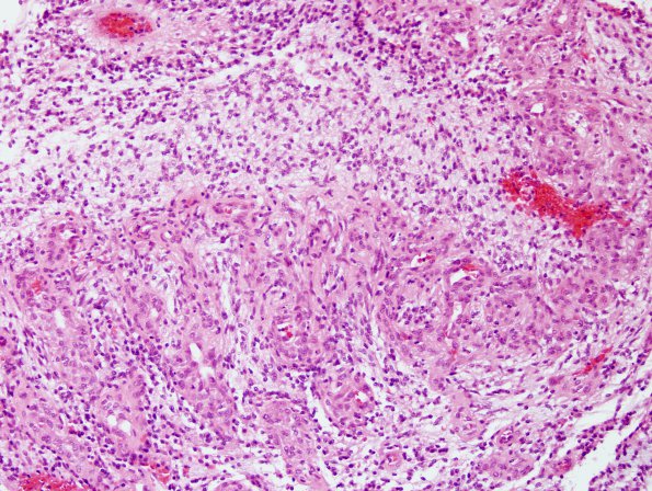
Sections from the original sellar/optic mass show a moderately cellular glial neoplasm in a predominantly myxoid background. There is mild nuclear pleomorphism, with the majority of tumor cells having oval to spindle-shaped nuclei with long thin cytoplasmic processes. In some areas, there are perivascular nuclear free zones, consistent with perivascular pseudorosettes. The tumor demonstrates extensive vascular proliferation which has a "glomeruloid" appearance in areas.

