Table of Contents
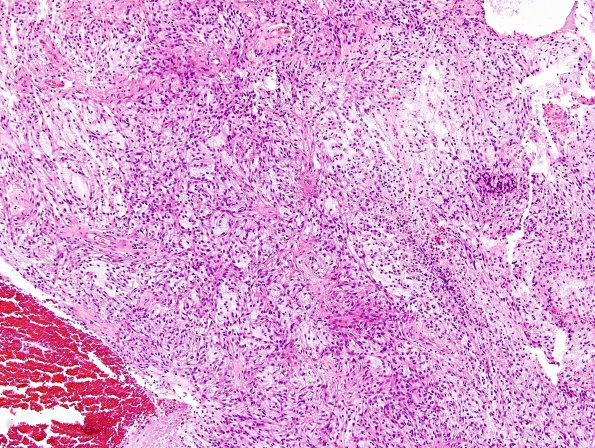
Washington University Experience | NEOPLASMS (GLIAL) | Astrocytoma, pilomyxoid | 5A1 Astrocytoma, pilocytic with pilomyxoid features (Case 5) H&E 7.jpg
Case 5 History ---- The patient is a 5 year old boy with two-and-a-half month history of vomiting and severe headaches. MRI showed a large posterior fossa mass with peripheral enhancement. Operative procedure: Resection. ---- 5A1-6 Sections reveal a moderately cellular solid and infiltrative appearing astrocytic neoplasm. In most areas, the tumor contains a loose mucin-rich stromal background and displays piloid cells with long thin eosinophilic cytoplasmic processes. Microcystic spaces are also evident in some areas and some foci display the classic biphasic appearance of pilocytic astrocytoma, including more compact regions. Only rare Rosenthal fibers and eosinophilic granular bodies are seen. Mitotic figures are hard to find, but there are large zones of tumor necrosis often associated with intravascular microthrombi. Vague perivascular pseudorosette-like structures are seen focally.

