Table of Contents
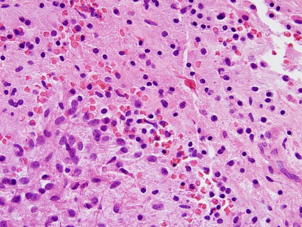
Washington University Experience | NEOPLASMS (GLIAL) | Astrocytoma, pilomyxoid | 5A6 Astrocytoma, pilocytic with pilomyxoid features (Case 5) H&E 8.jpg
Sections reveal a moderately cellular solid and infiltrative appearing astrocytic neoplasm. In most areas, the tumor contains a loose mucin-rich stromal background and displays piloid cells with long thin eosinophilic cytoplasmic processes. ---- Other stains (not shown) A histochemical stain for PAS with diastase highlights rare eosinophilic granular bodies. There is strong and widespread immunoreactivity for GFAP, highlighting the long piloid processes of the tumor cells. This stain also shows rare perivascular pseudorosette-like structures with processes radiating towards central blood vessels. The Ki-67 labeling index is generally low but shows increased activity focally. Much of this staining is seen within endothelial cells, but some of it appears to be in tumor cells as well.

