Table of Contents
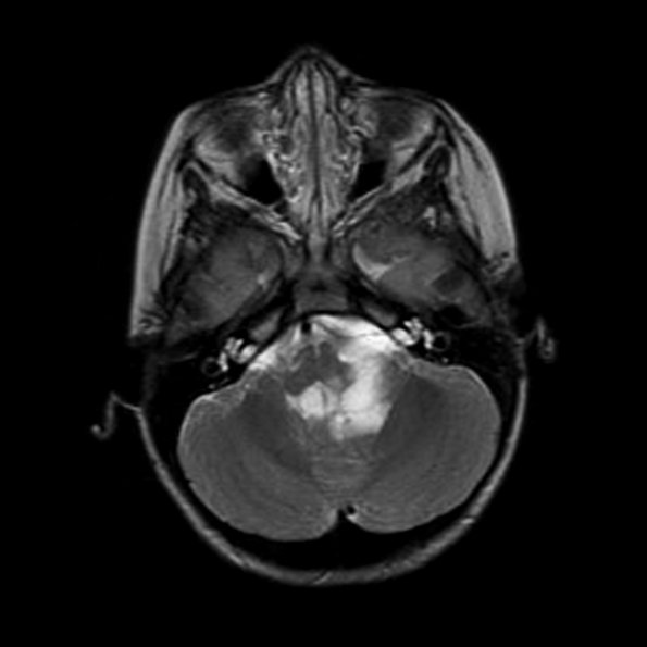
Washington University Experience | NEOPLASMS (GLIAL) | Astrocytoma, pilomyxoid | 6A4 Astrocytoma, pilomyxoid & PA (Case 6) MRI-T2
However, the mass was hyperintense on T2-weighted (6A4) scans. T2 studies revealed a heterogeneously enhancing left cerebellar/fourth ventricular mass, as well as two inferior spinal masses, suspicious for drop metastases. Intraoperatively, the surgeon observed "sugar coating" on the cerebellar surface, suggestive of subarachnoid spread.

