Table of Contents
Washington University Experience | NEOPLASMS (GLIAL) | Astrocytoma, pilomyxoid | 6B6 Astrocytoma, pilomyxoid & PA (Case 6) 5.jpg
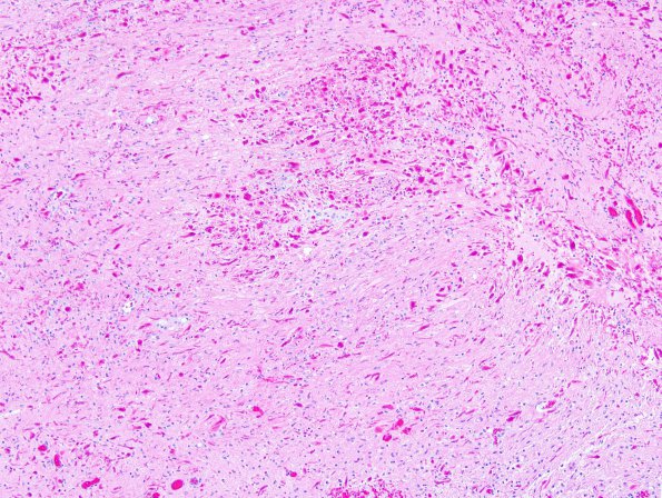
Sections reveal a moderately cellular solid and infiltrative neoplasm involving the cerebellum and overlying subarachnoid space. The tumor has mixed morphologic patterns. In some areas, it resembles pilomyxoid astrocytoma, containing a monomorphic population of thin spindled cells with a mucin-rich background and a prominent angiocentric growth pattern. The latter component resembles the pseudorosettes encountered in ependymal neoplasms. In these regions, Rosenthal fibers and eosinophilic granular bodies are not evident. In other areas, the tumor resembles a conventional pilocytic astrocytoma and is composed predominantly of the dense or compact element with elongate piloid cells, containing variably atypical nuclei and long, thin cytoplasmic processes. This component has exuberant Rosenthal fiber formation.

