Table of Contents
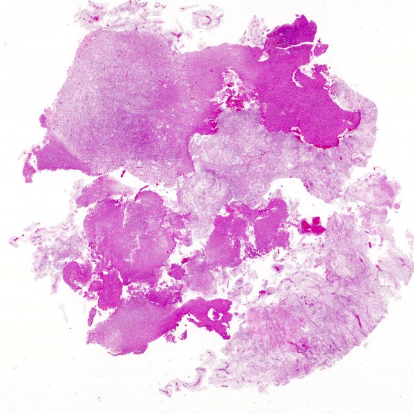
Washington University Experience | NEOPLASMS (GLIAL) | Astrocytoma, pilomyxoid | 7A1 Astrocytoma, pilomyxoid (Case 7) H&E whole mount
Case 7 History ---- The patient is a 1 year old boy with a 3 month history of shaking and vomiting. Imaging studies revealed a contrast enhancing mass involving the third ventricular and suprasellar region. ---- 7A1-7 Sections reveal a hypercellular glial neoplasm with mild pleomorphism. The majority of the tumor has a loose microcystic background, although focally, a more compact arrangement is also seen. The majority of tumor cells contain oval bland nuclei with long thin "piloid" cytoplasmic processes. In some areas, the tumor cells are arranged around blood vessels with perivascular nuclear-free zones reminiscent of perivascular pseudorosettes. Occasional mitotic figures are seen. There is no definite evidence of microvascular proliferation and only a small region of early necrosis is seen.

