Table of Contents
Washington University Experience | NEOPLASMS (GLIAL) | Astrocytoma, pilomyxoid | 8B1 Astrocytoma, Pilomyxoid (Case 8) H&E 3.jpg
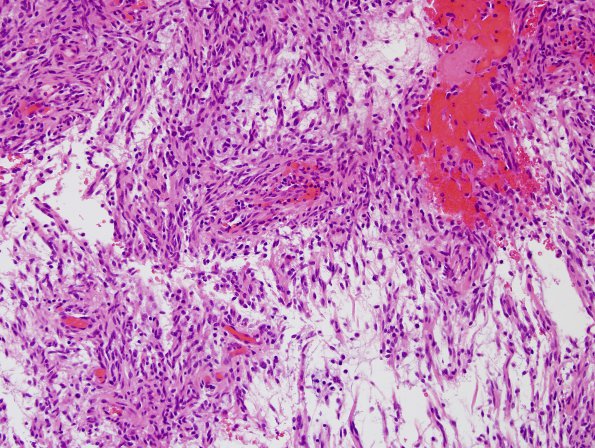
8B1-3 Specimen shows a moderately cellular astrocytic neoplasm composed of cytologically bland fusiform to spindle shaped with long eosinophilic cytoplasmic processes (piloid). There are few interspersed myxoid areas that appear hypocellular. Focally, there is a subtle indication of perivascular pseudorosettes. In addition, a few eosinophilic granular bodies are noted although Rosenthal fibers are not present, further supported by PAS with diastase stain. Rare mitoses are seen. However, areas of necrosis or microvascular proliferation are not seen.

