Table of Contents
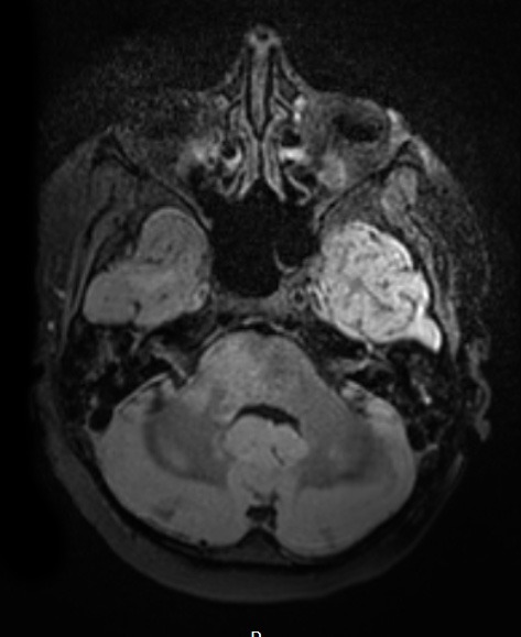
Washington University Experience | NEOPLASMS (GLIAL) | Diffuse midline glioma, H3 K27-altered | 15A3 DMG Bx (Case 15) T2W
This T2-weighted scan with administered contrast shows the involvement of the brainstem by the tumor. The hyperintensity of the left temporal lobe reflects terminal hypoxic/ischemic insult.

