Table of Contents
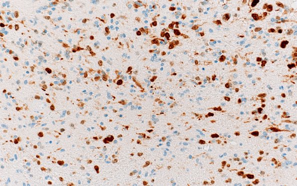
Washington University Experience | NEOPLASMS (GLIAL) | Diffuse midline glioma, H3 K27-altered | 2G DIPG (Case 2) N8 H3K27M N9 Ki67 40X 2
Ki67 highlights a proliferation index of approximately 30% in the most cellular areas of the neoplasm. ---- Not shown: There are patches of leptomeningeal involvement by tumor in the cerebral cortex, cerebellum, brainstem and spinal cord including the lumbosacral nerve roots. There is also subarachnoid hemorrhage associated with the tumor. Fibrin thrombi are noted in the leptomeningeal vessels. Multiple areas of neocortex show signs of acute and subacute hypoxic-ischemic injury, including hypereosinophilic necrotic neurons, microvacuolated parenchyma, and associated hemorrhage.

