Table of Contents
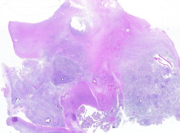
Washington University Experience | NEOPLASMS (GLIAL) | Diffuse midline glioma, H3 K27-altered | 3B1 DMG (Case 3) N14
3B1,2 Sections of the left thalamic mass shows a high-grade glioma. The tumor cells are pleomorphic, with atypical, hyperchromatic nuclei, prominent nucleoli, and moderate amounts of eosinophilic cytoplasm. Many tumor cells have large, bizarre nuclei, and frequent multinucleation. Some tumor cells have a short spindled morphology and others have a giant cell phenotype. Mitoses, including atypical forms, are numerous. Microvascular proliferation is robust.

