Table of Contents
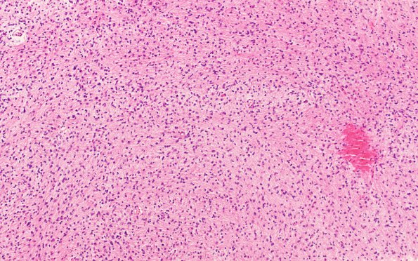
Washington University Experience | NEOPLASMS (GLIAL) | Diffuse midline glioma, H3 K27-altered | 4B1 Diffuse midline glioma, (Case 4) H&E 1
4B1-3 Routine H&E stained sections shows hypercellular brain parenchyma consisting of a diffusely infiltrating glial neoplasm, composed of tumor cells with indistinct cytoplasm with elongated atypical nuclei featuring hyperchromasia, coarse chromatin, and irregular nuclear contours. Mitotic figures are readily identified. There are focal areas of incipient necrosis but no evidence of microvascular proliferation.

