Table of Contents
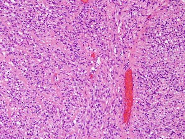
Washington University Experience | NEOPLASMS (GLIAL) | Ependymoma, clear cell | 4B1 Ependymoma, extra-axia, clear & tanycyticl (Case 4) H&E 5
4B1-4 The tumor is composed of a cellular proliferation of elements with spindled morphology and round, ovoid, and spindle-shaped nuclei. Most of the cells have elongated, eosinophilic processes with indistinct cell borders. The nuclei are rather bland-appearing, predominantly ovoid in shape with mild contour irregularities and smooth, finely granular to vesicular chromatin; scattered atypical forms are identified. Scattered vessels show perivascular chronic inflammation composed of lymphocytes and plasma cells and hemosiderin-laden macrophages. Additionally, there are areas of tanycytic as well as focal clear cell features. There is a relative paucity of ependymal rosettes and pseudorosettes. Mitotic figures are identified at a rate of 2-3 per 10 high-powered fields. No microvascular proliferation or necrosis is seen.

