Table of Contents
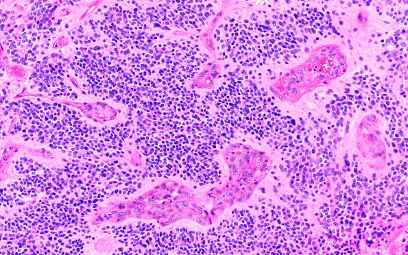
Washington University Experience | NEOPLASMS (GLIAL) | Ependymoma, clear cell | 7A1 Ependymoma, anaplastic, clear cell (Case 7) H&E 20X
Case 7 History ---- The patient is an 11 year old boy with a 6.5 cm, partially cystic, partially calcified mass with peripheral contrast enhancement in the left frontal lobe. Diagnosis: Anaplastic ependymoma with clear cell features, WHO 3. ---- 7A1,2 Microscopically, sections reveal a demarcated cellular neoplasm with scattered calcifications. There is mild to moderate nuclear pleomorphism, with the majority of tumor cells containing round to oval nuclei with inconspicuous nucleoli and variable quantities of clear to amphophilic cytoplasm. In many areas, there are well formed perivascular pseudorosettes and a few true rosettes. There is extensive microvascular proliferation and foci of infarct-like necrosis are similarly prominent. Mitotic figures are numerous, reaching up to 6 mitoses per 10 high power fields.

