Table of Contents
Washington University Experience | NEOPLASMS (GLIAL) | Ependymoma, myxopapillary | 10A2 Ependymoma, myxopapillary (Case 10) T1 W2 - Copy - Copy copy
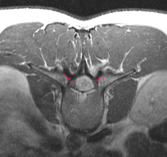
A classic boxcar appearance of this T1-weighted contrast administered image is also shown in axial section surrounded by the roots of the cauda equina (arrows).

