Table of Contents
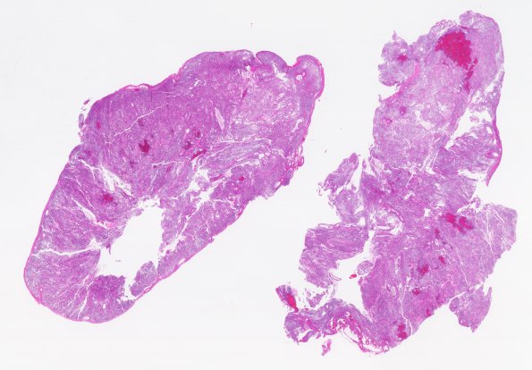
Washington University Experience | NEOPLASMS (GLIAL) | Ependymoma, myxopapillary | 10B1 Ependymoma, myxopapillary (Case 10) H&E 9
10B1-4 Sections show a cellular tumor composed of perivascular pseudorosettes and short fascicles of spindled tumor cells with intervening myxoid material. The vessels within the tumor appear to have a thickened collagenous rim and a very rare examples shows rudimentary endothelial proliferation. There are several areas of tumor necrosis along with hemosiderin and hematoidin pigment deposition. Most tumor nuclei are elongated with dense chromatin and only occasional pleomorphism is seen. The mitotic rate is generally low overall in the tumor, however, up to 4 mitoses per 10 high powered fields can be seen in some areas.

