Table of Contents
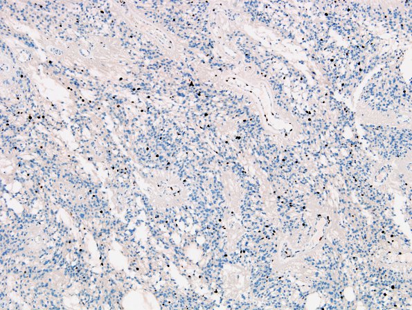
Washington University Experience | NEOPLASMS (GLIAL) | Ependymoma, myxopapillary | 10E Ependymoma, myxopapillary (Case 10) MIB 2
10E Proliferation as measured by a Ki-67 labeling index shows 6.3% of cells are positive, however, there is significant labeling of endothelial cells which contribute to the overall count. ---- Comment: The morphologic and immunohistochemical features of this tumor are most consistent with a myxopapillary ependymoma, which was called MPE with increased proliferation index. A few foci of necrosis are also encountered as well as a few patches of endothelial proliferation. It does not have regional hypercellularity or reduced mucin but does have ≥ 2 mitoses/mm2, minimal microvascular proliferation, and spontaneous necrosis. It is not clearly anaplastic MPE to me, which is ungraded in the WHO 2021. The patient followup has been uneventful with no active growth in 7 years.

