Table of Contents
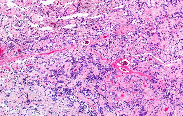
Washington University Experience | NEOPLASMS (GLIAL) | Ependymoma, myxopapillary | 11A1 Ependymoma, myxopapillary (Case 11) H&E 5
Case 11 History ---- The patient is a 68-year-old woman with thoracolumbar junction pain. An MRI scan revealed tight stenosis at L1-2 associated with retrolisthesis and disc degeneration focally at the L1-2 level. She underwent L1 Gill procedure for decompression of the neural element and L1-2 post lumbar interbody fusion on 12/12/2018. An intradural extramedullary lesion was identified in the cauda equina and was resected. ---- 11A1-5 There is focal papillary architecture with cuboidal to elongated cells radially arranged along strongly hyalinized fibrovascular core. The tumor cells have round to oval nuclei with delicate chromatin and inconspicuous nucleoli. Microcysts with mucin pools are also seen. Mitotic activity is not increased. (H&E)

