Table of Contents
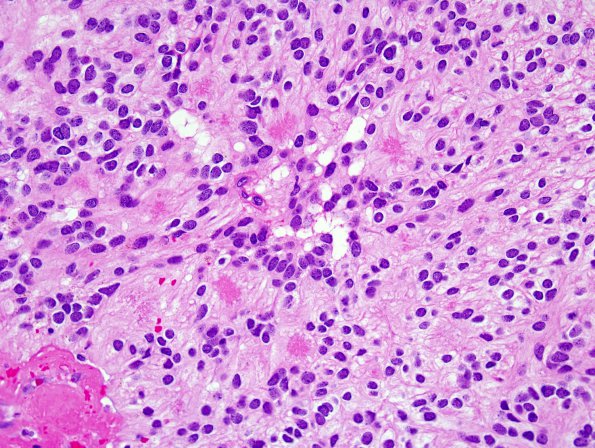
Washington University Experience | NEOPLASMS (GLIAL) | Ependymoma, myxopapillary | 13A2 Ependymoma, myxopapillary (Case 13) H&E 5.jpg
Sections of the neurosurgical specimen consist of a moderately cellular neoplasm arranged in a prominent papillary pattern. The tumor cells are arranged in these papillary structures in a radiating pattern surrounding centrally placed markedly thickened and hyalinized blood vessels (also highlighted by trichrome stain). The tumor cells are bland, short spindled to epithelioid, have eosinophilic cytoplasm and contain nuclei with dispersed chromatin pattern. True rosettes are not seen. Mitoses are present, albeit rare.

