Table of Contents
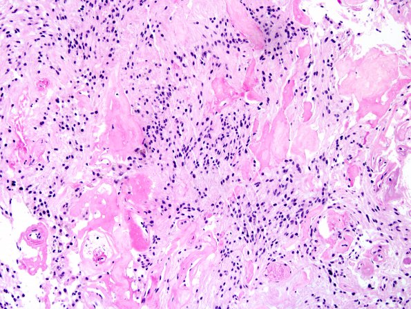
Washington University Experience | NEOPLASMS (GLIAL) | Ependymoma, myxopapillary | 15A4 Ependymoma, myxopapillary (Case 15) H&E 8.jpg
Sections show a proliferation of spindled to cuboidal cells with round-to-ovoid nuclei, elongated eosinophilic processes, and occasional, moderate nuclear atypia. Papillary architecture is somewhat inconspicuous. Many of the vessels appear hyalinized. There are focal areas of myxoid background stroma, although many areas are solid. Microvascular proliferation and necrosis are not identified. Small round, spiculated "collagen balloons" are noted.

