Table of Contents
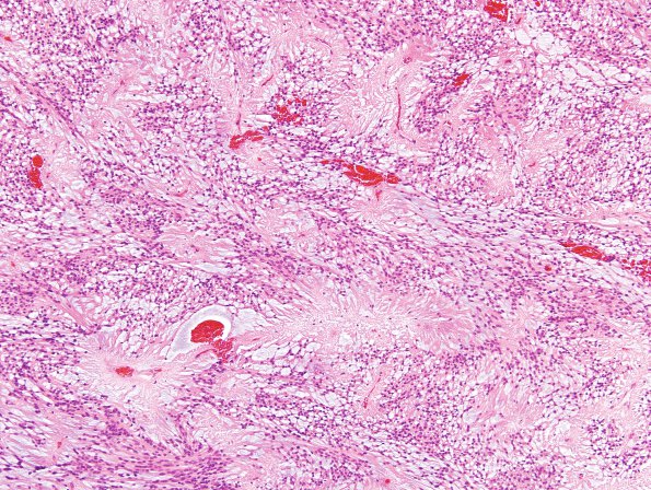
Washington University Experience | NEOPLASMS (GLIAL) | Ependymoma, myxopapillary | 16B1 Ependymoma, myxopapillary (Case 16) H&E 3
16B1 Histological sections of the L1-L2 spinal cord mass show an encapsulated glial neoplasm composed of long, bipolar tumor cells with bland, round-to-oval nuclei, finely speckled chromatin, small basophilic nucleoli, and eosinophilic cytoplasm with poorly-defined intercellular borders. These cells are radially arranged into striking and abundant perivascular pseudorosettes, and cell processes are often separated from each other by abundant small pools of bubbly blue mucin. Papillary architecture is not present. Mitotic figures are not observed.

