Table of Contents
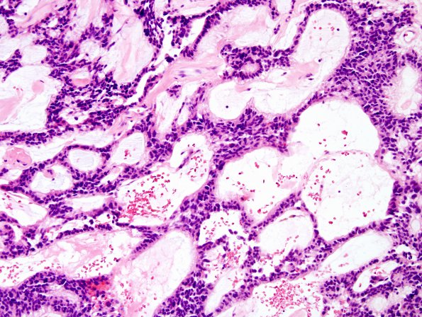
Washington University Experience | NEOPLASMS (GLIAL) | Ependymoma, myxopapillary | 17B Ependymoma, myxopapillary (Case 17) H&E 2
Sections from both the specimens demonstrate a moderately cellular neoplasm with large number of markedly hyalinized blood vessels and focal areas of hemosiderin deposition. The tumor cells are organized in a radiating pattern surrounding centrally placed hyalinized blood vessels and are short spindled to epithelioid, containing eosinophilic cytoplasm. In some areas, the tumor becomes more cellular and in these foci, the nuclei show significant degenerative atypia. The tumor has a prominent papillary architecture with scattered perivascular pseudo-rosettes. Many of these papillae have an intermediate layer of myxoid (basophilic mucinous) degeneration in their walls, focally, forming microcysts. Mitoses are difficult to find and microvascular proliferation and/or necrosis are not seen.

