Table of Contents
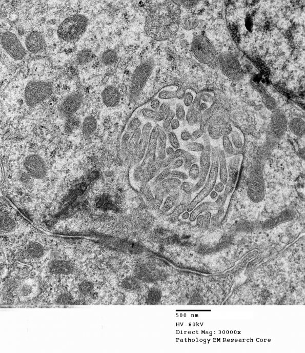
Washington University Experience | NEOPLASMS (GLIAL) | Ependymoma, myxopapillary | 18G5 Ependymoma, myxopapillary (Case 18)_013 - Copy - Copy
Higher magnification of image #18G4 (electron micrograph) ---- Based on histomorphologic features and immunostaining profile, this neoplasm is most consistent with an ependymal neoplasm with extensive hemorrhage and necrosis. However, this neoplasm is difficult to precisely classify due to extensive hemorrhage, necrosis and lack of a microcystic, mucinous background or perivascular pseudorosettes. However, the papillary architecture and location suggest a myxopapillary ependymoma, WHO grade 2.

