Table of Contents
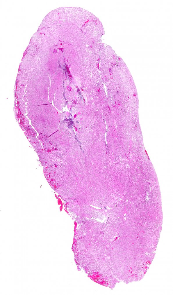
Washington University Experience | NEOPLASMS (GLIAL) | Ependymoma, myxopapillary | 19A1 MPE (Case 19) H&E WM
Case 19 History ---- The patient is a 33 year old woman who was involved in a motor vehicle accident three years ago. Six months ago, following a fall, she had a constant ache in her lower back that radiated to the right leg. ---- Imaging studies showed a hyperintense signal abnormality that is enhancing behind the body of L3 and L4 vertebrae. Clinical diagnosis: Intradural tumor. Operative procedure: Resection. ---- 19A1,2 Sections of the "intradural tumor" show a myxopapillary ependymoma. The neoplasm is composed of clusters or sheets of uniform cells with round to oval nuclei and finely granular chromatin pattern in a mucinous background. There is focal pleomorphism with scattered large nuclei. A few mitotic figures are found. There is no evidence of necrosis or vascular proliferation. There is focal true rosette formation and a focally prominent fibrillary background. In other areas, ependymal canal and pseudorosette formation are present.

