Table of Contents
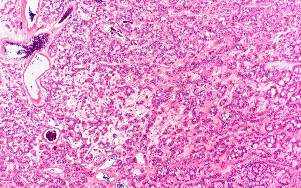
Washington University Experience | NEOPLASMS (GLIAL) | Ependymoma, myxopapillary | 19A2 MPE (Case 19) 4X H&E
Sections of the intradural tumor show a myxopapillary ependymoma. The neoplasm is composed of clusters or sheets of uniform cells with round to oval nuclei and finely granular chromatin pattern in a mucinous background. There is focal true rosette formation and a focally prominent fibrillary background. In other areas, ependymal canal and pseudo rosette formation are present.

