Table of Contents
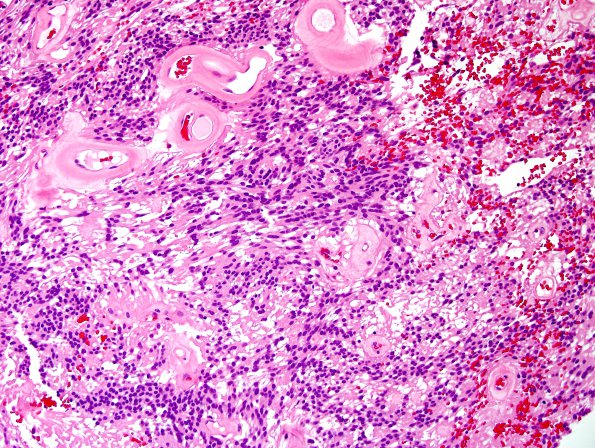
Washington University Experience | NEOPLASMS (GLIAL) | Ependymoma, myxopapillary | 1B2 Ependymoma, myxopapillary (Case 1) H&E 1.jpg
Sections of the resected spinal cord tumor show a low-grade primary neoplasm. Much of the tumor displays low cellularity, with cells arranged into clusters of nuclei separated by nucleus-free zones and occasional mucin-filled microcystic spaces. Nuclear cytology is bland and uniform and delicate hair-like cytoplasmic processes extend from opposite ends to impart bipolar morphology. Vessels display extensive hyalinization, and perivascular pseudorosette formation. Mitotic activity is absent. Although there are large areas of necrosis, this does not indicate malignancy in a tumor that lacks additional high grade features. Ancillary workup was performed to further characterize this neoplasm.

