Table of Contents
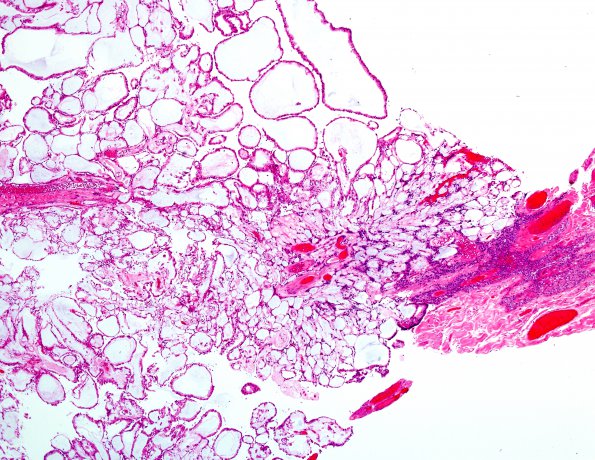
Washington University Experience | NEOPLASMS (GLIAL) | Ependymoma, myxopapillary | 20A2 Ependymoma, myxopapillary (Case 20) H&E 1.jpg
Section of the intradural lesion shows thin walled micropapillary bundles surrounded by a single layer of epithelium that varies from low cuboidal to columnar. The cores of the papillae contain variable amount of a mucinous/myxoid matrix.

