Table of Contents
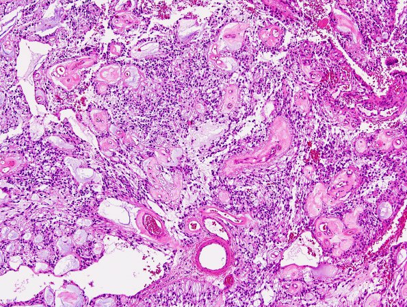
Washington University Experience | NEOPLASMS (GLIAL) | Ependymoma, myxopapillary | 2C2 Ependymoma, myxopapillary (Case 2) H&E 1.jpg
Hematoxylin and eosin stained sections of the intradural mass show a well circumscribed tumor with a prominent myxoid background. The tumor cells are oval to elongated, some of which are arranged around thick, hyalinized blood vessels. Areas of microcysts filled with mucinous material are also present. Focally present in the tumor are areas of papillary growth with radially arranged tumor cells. Only rare mitoses are seen (< 1/10HPF), and there are small foci with bland necrosis.

