Table of Contents
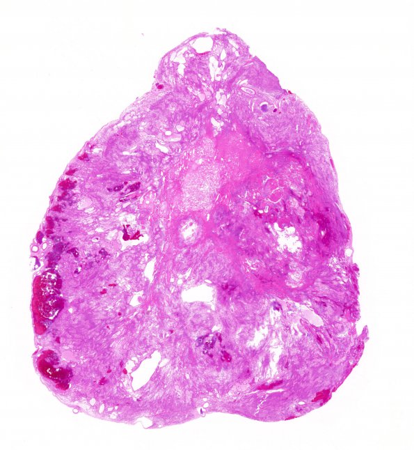
Washington University Experience | NEOPLASMS (GLIAL) | Ependymoma, myxopapillary | 3B1 Ependymoma, myxopapillary (Case 3) 1 H&E WM
3B1-6 H&E stained sections show multiple fragments of a well-circumscribed, partially encapsulated, ependymal neoplasm. There are some areas of bland infarct-type necrosis. There are patchy areas of microcysts that contain extracellular pale basophilic material. There are abundant thickened and hyalinized vessels. Surrounding these vessels are relatively anuclear zones, which are then surrounded by tumor cells with an eosinophilic fibrillary background. The tumor cells are without distinct cell-cell borders and the nuclei are round to oval with finely speckled chromatin. Mitotic figures are not readily identified.

