Table of Contents
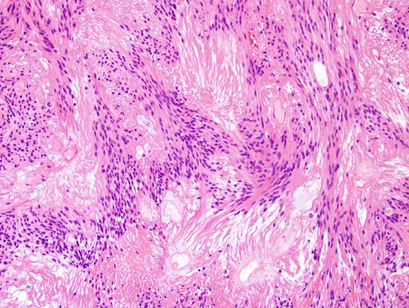
Washington University Experience | NEOPLASMS (GLIAL) | Ependymoma, myxopapillary | 3B5 Ependymoma, myxopapillary (Case 3) H&E 8.jpg
Surrounding these vessels are relatively anuclear zones, which are then surrounded by tumor cells with an eosinophilic fibrillary background. The tumor cells are without distinct cell-cell borders and the nuclei are round to oval with finely speckled chromatin. Mitotic figures are not readily identified.

