Table of Contents
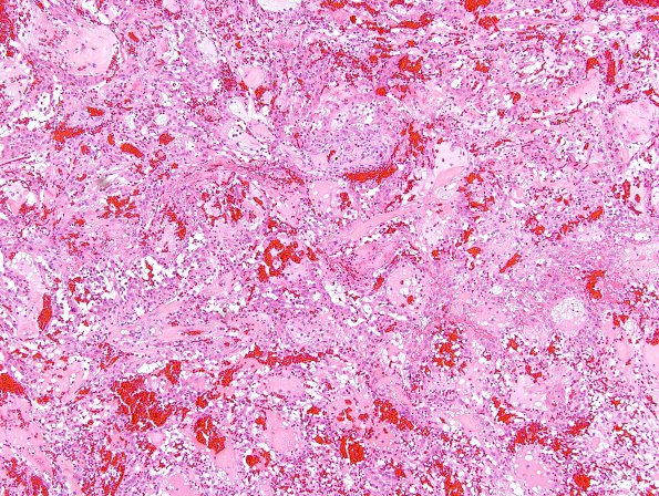
Washington University Experience | NEOPLASMS (GLIAL) | Ependymoma, myxopapillary | 6A2 Ependymoma, myxopapillary (Case 6) H&E 6
Sections of the conus tumor show a well circumscribed mass. The capsule is thin and focally disrupted. The tumor itself consists of plump, almost epithelioid cells with abundant pink cytoplasm and round to oval nuclei, with prominent nucleoli. These form an admixture of more solid areas and areas which form pseudo-acini filled with colloid-like myxoid material. There are also prominent vascular channels and blood vessels. Abundant hemosiderin-laden macrophages are scattered throughout, which are mainly concentrated at the periphery.

