Table of Contents
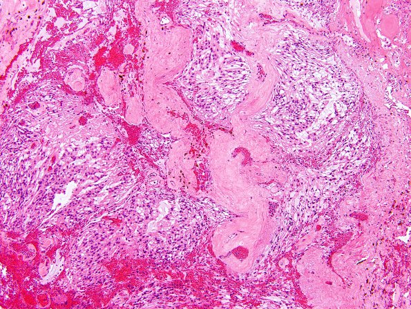
Washington University Experience | NEOPLASMS (GLIAL) | Ependymoma, myxopapillary | 7A1 Ependymoma, myxopapillary, anaplastic fx (Case 7) - H&E representative 4 - low powerA.jpg
Case 7 History ----The patient is a 57-year-old man who was status post partial resection of a lumbar myxopapillary ependymoma, WHO grade I, at age 45 with subsequent radiation therapy. ---- 7A1,2 The original neoplasm was composed of infiltrative sheets and clusters of ependymal cells embedded in a myxoid stroma. Although occasionally the tumor cells form solid arrays with abundant, elongate glial processes, much of the tumor shows a microcystic pattern of growth. Only mild cytologic atypia is present and the mitotic rate is very low. No areas of tumor necrosis or vascular proliferation are present. Islands of neoplastic tissue are separated by a highly vascular connective tissue in which abundant hemosiderin and clotted blood are present.

