Table of Contents
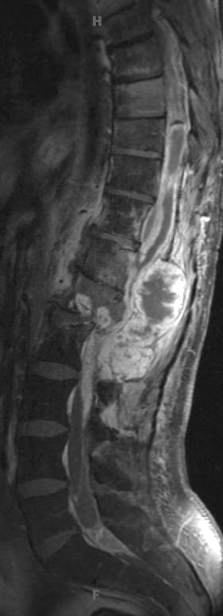
Washington University Experience | NEOPLASMS (GLIAL) | Ependymoma, myxopapillary | 7B2 Ependymoma, myxopapillary, anaplastic fx (Case 7) -MRI T1post - Copy
MRIs are shown consisting of T1 weighted pre-contrast (7B1), T1-weighted post contrast (7B2) and T2 (7B3). There are central areas of fluid signal intensity and decreased enhancement consistent with cystic change or central necrosis. Focal abnormal signal and enhancement are noted in the thecal sac, suggestive of drop metastasis.

