Table of Contents
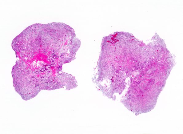
Washington University Experience | NEOPLASMS (GLIAL) | Ependymoma, myxopapillary | 8A1 Ependymoma, myxopapillary (Case 8) 1 H&E WM
Case 8 History ---- The patient is a 60-year-old woman with progressive right leg dysfunction. MRI of the lumbar spine showed a 7 mm focus of enhancement within the thecal sac at the L1 level. Radiologic impression: Intradural schwannoma. Operative procedure: Posterior thoraco-lumbar laminectomy with resection of an intradural extramedullary mass. ---- 8A1-4 Sections showed a low-grade glial neoplasm consisting of neoplastic cells with a spindled appearance and a perivascular distribution. The neoplastic cells have ovoid to round nuclei with homogenously basophilic staining chromatin. The vasculature is single-layered and shows prominent hyalinization. In many areas, the neoplastic cells form loose fascicles separated by a pale blue myxoid stroma. Mitotic activity is not appreciated.

