Table of Contents
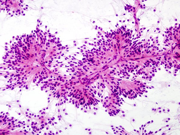
Washington University Experience | NEOPLASMS (GLIAL) | Ependymoma, myxopapillary | 9A4 Ependymoma, myxopapillary (Case 9) Smear 8
Sections from the neurosurgical specimen consist of a moderately cellular neoplasm arranged in a prominent papillary pattern. Many of the papillae have myxoid degeneration in their center. The tumor cells are arranged in a radiating pattern surrounding centrally placed markedly hyalinized blood vessels and often, an intermediate layer of mucoid degeneration. The tumor cells are bland, short spindled to epithelioid, have eosinophilic cytoplasm and contain nuclei with dispersed chromatin pattern. True rosettes are not seen. Mitoses are present, albeit rare. Patchy microvascular proliferation and a minute region of necrosis are also seen. The overall morphological findings are consistent with myxopapillary ependymoma, WHO grade I (now WHO Grade 2).

