Table of Contents
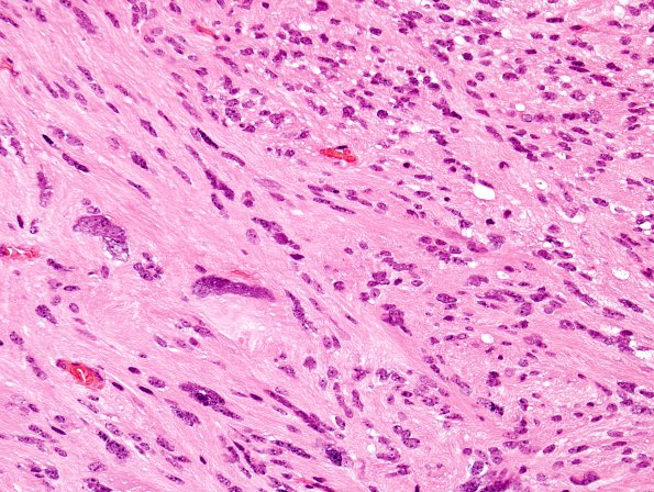
Washington University Experience | NEOPLASMS (GLIAL) | Ependymoma, tanycytic | 2A2 Ependymoma, tanycytic (Case 2) H&E 1 (2)
2A2-5 Scattered throughout the lesion are numerous tumor cells displaying enlarged, bizarre shaped hyperchromatic nuclei. Vague perivascular pseudorosette formations are detected, though no true ependymal canals or frank rosettes are present. Occasional mitotic figures are detected. There is no evidence of necrosis or endothelial hyperplasia.

