Table of Contents
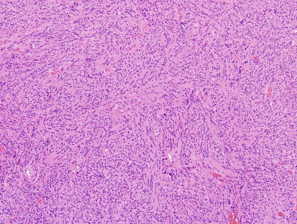
Washington University Experience | NEOPLASMS (GLIAL) | Ependymoma, tanycytic | 5A1 Ependymoma, tanycytic (Case 5) H&E 1
Case 5 History ---- The patient is a 47 year old woman with an approximately five year history of low back pain. Within the last six months, this progressed to right lower extremity pain. MRI studies revealed an intrathecal lumbar spinal cord mass at the L1-2 level. The imaging differential diagnosis included ependymoma, schwannoma, and meningioma. ---- 5A1-3 Sections reveal a solid-appearing spindle cell neoplasm with moderate cellularity and in a “single file” growth pattern. There is mild nuclear pleomorphism, with the majority of tumor nuclei being oval with delicate chromatin and abundant eosinophilic cytoplasm. Perivascular pseudorosettes are infrequent, if present at all. Mitotic figures are rare and there is no definite evidence of microvascular proliferation or tumor necrosis. Scattered bizarre nuclei are also evident, likely degenerative in nature.

